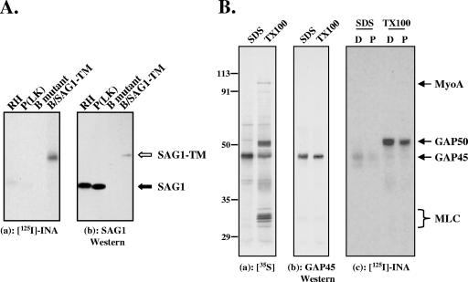
FIG. 2.
INA preferentially labels non-GPI-linked integral membrane proteins of the pellicle. (A) (a) SAG1 was immunoprecipitated from parasites labeled with INA through photosensitization and analyzed by SDS-PAGE/autoradiography. The parasite strains used were RH and P(LK) (wild-type strains), a SAG1-deficient P(LK) strain (B mutant), and the SAG1-deficient strain expressing a transmembrane version of SAG1 (B/SAG1-TM). (b) Corresponding Western blot with anti-SAG1 antibody. GPI-anchored SAG1 shows weak labeling with INA (panel a, filled arrow). In contrast, much smaller amounts of SAG1-TM [compare Western blotting signals of RH and P(LK) to that of B/SAG1-TM in panel b] show significantly more INA incorporation (panel a, open arrow). The lanes shown were from the same blot and were exposed and adjusted for contrast and brightness identically. (B) (a) Autoradiogram showing that immunoprecipitation of SDS-extracted 35S-labeled parasites with an anti-GAP45 antibody yields primarily GAP45 (SDS), whereas the entire glideosome complex can be immunoprecipitated with the same antibody under nondenaturing conditions (TX-100). The four major proteins in the immunoprecipitated complex are myoA, myosin light chain (MLC), GAP45, and GAP50 (11). (b) Corresponding Western blot with anti-GAP45. (c) Autoradiogram of the INA-labeled proteins immunoprecipitated with anti-GAP45 antibody shows that both GAP45 and GAP50 are labeled, through either direct or photosensitized INA activation, whereas neither myosin light chain nor myoA is detectably labeled. Substoichiometric amounts of myoA were transferred to the nitrocellulose in this particular experiment; other experiments in which the iodination profile of the immunoprecipitate was analyzed directly (i.e., without transfer for Western blotting) confirmed the lack of detectable INA incorporation into myoA (data not shown). D, direct activation; P, photosensitization. Numbers on the left indicate molecular mass in kDa.

