FIG. 6.
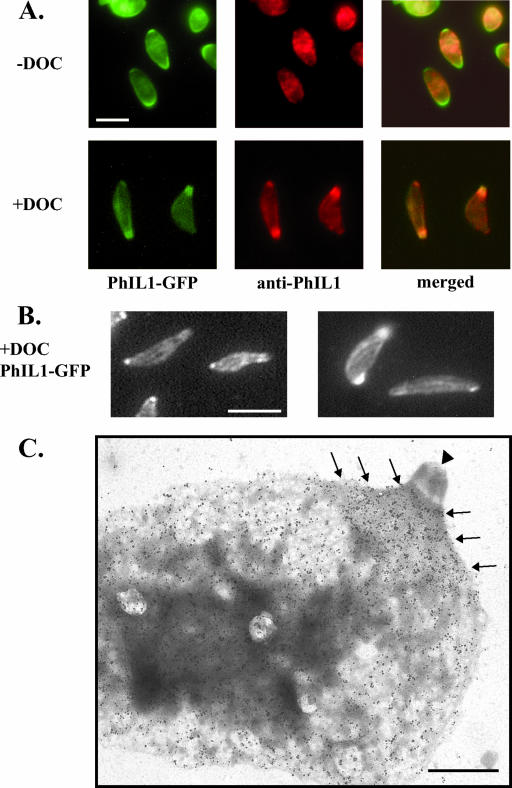
Localization of PhIL1 in DOC-extracted parasites. (A) In fixed, TX-100-permeabilized parasites expressing PhIL1-GFP (− DOC), both anti-PhIL1 (middle panels) and anti-GFP (not shown) localize to the parasite periphery by immunofluorescence microscopy, but neither shows the apical and posterior concentration seen by direct fluorescence microscopy of PhIL1-GFP (left panels). However, after extraction with deoxycholate (+ DOC), both antibodies label the apical and posterior ends. Bar, 5 μm. (B) In DOC-extracted parasites, PhIL1 is also found in spiraling “stripes” along the body of the parasite. Two independent examples of this labeling pattern are shown. Bar, 5 μm. (C) DOC-extracted PhIL1-GFP parasites were incubated with either preimmune sera or anti-PhIL1 antibody, followed by gold labeling and negative staining of the parasite “ghosts” with phosphotungstic acid. Very little labeling is seen in the controls labeled with preimmune serum (data not shown). Using anti-PhIL1, labeling is observed along the body of the parasite and in a concentrated “cape” (arrows) at the apical end, just basal to the conoid (arrowhead). No labeling of the conoid is seen. Labeling of the basal end of the parasite is also observed, although this labeling is less pronounced than that at the apical end (data not shown). Bar, 1 μm.

