FIG. 8.
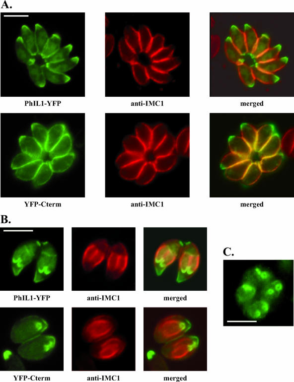
The conserved C terminus targets PhIL1 to the periphery and to the apex of developing daughter parasites during endodiogeny. (A) The C-terminal portion of PhIL1 (YFP-Cterm) localizes to the parasite periphery and is concentrated at the apical end. The distributions of PhIL1-YFP and YFP-Cterm (direct fluorescence) are shown, relative to IMC1 (indirect immunofluorescence). In contrast, an N-terminal YFP fusion of PhIL1 does not target itself to the periphery of the parasite, although it is found at the apical end (data not shown). (B) During parasite replication, where daughter parasites form within the mother, full-length PhIL1-YFP localizes to the apical ends of the forming daughters, anterior to the gap in IMC1. YFP-Cterm is similarly targeted to daughter parasites. (C) When daughter parasites are oriented with their apical end towards the reader, it is clear that the gap in PhIL1 apical fluorescence (here PhIL1-GFP) corresponds to a tight circle. All bars, 2.5 μm.

