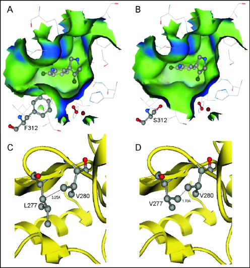
FIG. 2.
(A and B) Molecular surfaces in the substrate binding pockets of wild-type PrnD (A) and the F312S mutant (B). Blue and green indicate hydrophilic and hydrophobic surfaces, respectively. (C and D) The intermolecular distances between V280 and L277 in wild-type PrnD (C) and V277 in the L277V mutant (D) are 3.50 and 1.70 Å, respectively. The molecular surfaces and intermolecular distances are the result of modeling.

