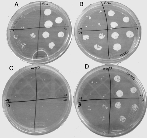
FIG. 4.
Colony appearance of wild-type (A and B) and ΔmshD M. tuberculosis (C and D) cells grown on Middlebrook 7H9 plates supplemented with ADS (A and C) or OADC (B and D). Cells were plated at dilutions of 10−1 (upper right quadrant), 10−2 (lower right quadrant), 10−3 (lower left quadrant), and 10−4 (upper left quadrant). Plates were photographed after 4 weeks of incubation.

