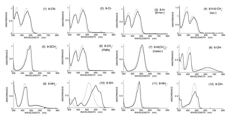
Figure 1.
Absorption spectra of a series of FMN analogs (dotted lines) and the reconstituted lactate oxidase with these analogs (solid lines) in 0.1 M phosphate buffer (pH 7.0) at room temperature. The peak intensity of each FMN analog in free solution was normalized as unity. The numberings of absorbance spectra are based on the order of the redox midpoint potential of FMN analogs as follows: 1, 8-CN-FMN; 2, 8-Cl-FMN; 3, 8-H-FMN (norflavin); 4, 8-H,6-CH3-FMN (isoflavin); 5, 8-CH3S-FMN; 6, FMN; 7, 8-(CH3)2N-FMN (roseoflavin); 8, 6-OH-FMN, 9, 6-NH2-FMN; 10, 8-SH-FMN; 11, 8-NH2-FMN; 12, 8-OH-FMN.

