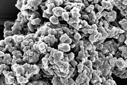
Fig. 1.
SEM image of dried NM pigment isolated from the SN region of human brain tissue. The image illustrates a commonly observed, large, amorphous aggregate (>10 μm in size) composed of smaller NM molecules, roughly spherical in shape and varying widely (200–500 nm) in diameter. The image is 6.75 μm × 4.50 μm.

