Fig. 1.
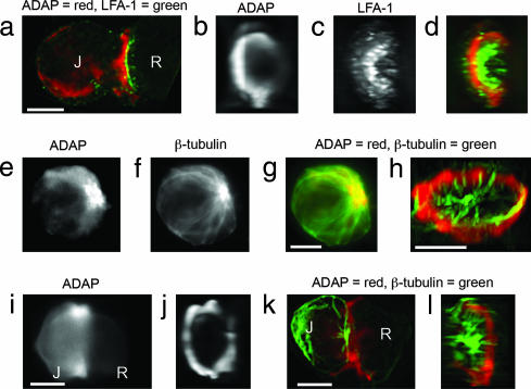
ADAP, LFA-1, and microtubules at the Jurkat immunological synapse. (a–d) SEE-coated Raji (R) cells conjugated to Jurkat (J) cells were fixed and immunostained for LFA-1 and ADAP. The z-axis stacks (256 images) were acquired for LFA-1, and ADAP fluorescence data were processed as described (3). A Jurkat–Raji pair immunostained for ADAP (red) and LFA-1 (green) is shown from a side view (a) and facing the synapse with separate panels for ADAP (b) and LFA-1 (c). A red–green overlay of the data from b and c is shown in d. Note that the ADAP and LFA-1 rings are separate and distinct. (e–g) Jurkat cells were immunostained for ADAP (e) and tubulin (f) with the red–green overlay shown in g (red, ADAP; green, tubulin). (h) SEE-coated Raji cells pretreated with colchicine to depolymerize microtubules were mixed with normal Jurkat cells and then immunostained for tubulin (green) and ADAP (red). Image stacks were acquired and processed as for a–d. A typical Jurkat–Raji pair shows microtubules projecting from the MTOC to the ADAP ring where the microtubules often closely follow its surface. A rotatable view is available as Movie 1. (i–j) Cells were prepared identically to the procedure in a–d except that Jurkat cells were pretreated with 10 μM colchicine to depolymerize the microtubules. The results show that the ADAP ring forms in the absence of an intact microtubule cytoskeleton. (k and l) Cells were prepared identically to the procedure in a–d except that JB2.7 (LFA-1-deficient) Jurkat cells were used in place of normal Jurkat cells. The results show that microtubules are associated with a typical ADAP ring in the absence of LFA-1. Data are representative of two to three independent experiments. (Scale bars: 5 μm.)

