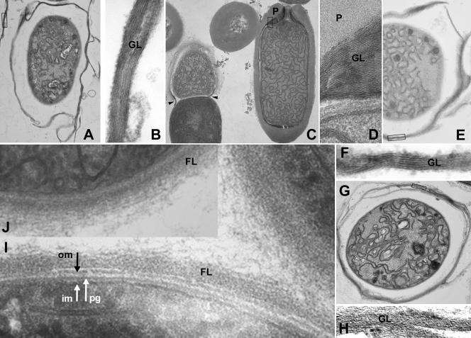
FIG. 1.
Electron microscopy of heterocyst sections from regulatory Fox− mutants. (A and B) alr0117 mutant FQ671. (C and D) all0187 mutant FQ1062, most of whose heterocysts have terminal pores that are unusually wide (C, arrowheads: the heterocyst on the right is more nearly normal in structure). (E and F) all2760 mutant FQ1641. (G and H) alr5348 mutant FQ1281. (I and J) alr1086 mutant FQ621. Panels B, D, F, and H, magnified from the boxed regions in panels A, C, E, and G, respectively, show glycolipid laminations. im, inner membrane; pg, peptidoglycan; om, outer membrane; GL, envelope glycolipid; P, envelope polysaccharide; FL, fibrous material. Magnifications (103): A, ×11; B, ×142; C, ×9.3; D, ×148; E, ×19; F, ×147; G, ×15; H, ×118; I, ×137; J, ×69.

