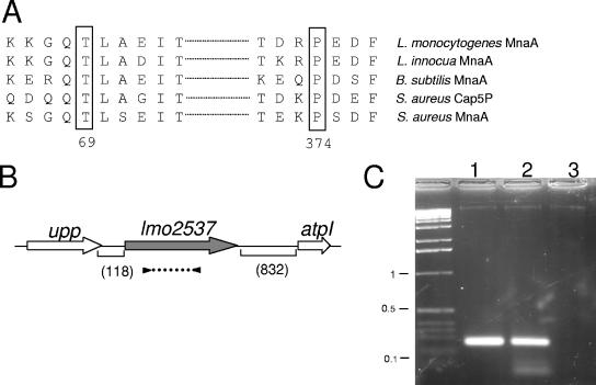
FIG. 1.
Schematic organization of the lmo2537 locus. (A) Alignment of the B. subtilis MnaA domains comprising T69 and P374 with their counterparts in S. aureus, L. monocytogenes, and L. innocua. (B) lmo2537 gene diagram. Arrows indicate the orientation and approximate sizes of the open reading frames. Parenthetic numbers give the sizes (in base pairs) of the intergenic regions flanking lmo2537. upp, uracil phosphoribosyltransferase; lmo2537, UDP-GlcNAc 2-epimerase; atpI, ATP synthase subunit I. The dotted line flanked by black triangles below lmo2537 indicates the positions of the primers used in the RT-PCR analysis. (C) Tris-acetate-EDTA-agarose gel electrophoresis of transcripts amplified by RT-PCR. Numbers on the left correspond to the sizes (in kilobases) on the DNA ladder. Lanes: 1, PCR on control DNA; 2, RT-PCR plus RNA; 3, RT-PCR without RT.

