
Figure 3.
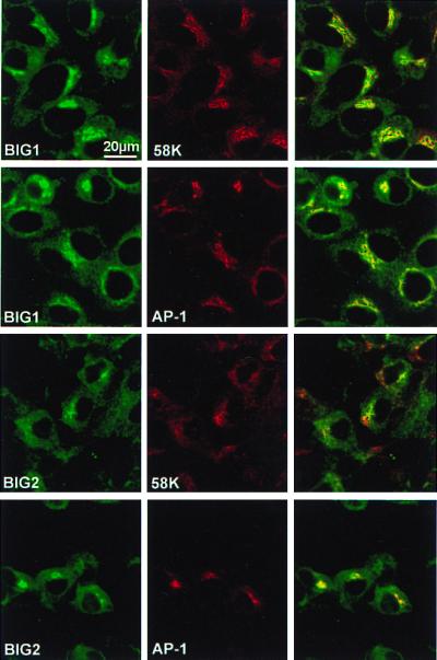
Golgi localization of BIG1 and BIG2. HepG2 cells were reacted with rabbit anti-BIG1 or anti-BIG2 IgG and mouse anti-58K or -AP-1 antibodies, as indicated, followed by FITC-labeled anti-rabbit IgG and Texas Red-labeled anti-mouse IgG. In the third panel of each row, images of the preceding two panels are superimposed. Observations were replicated with other samples of cells, five times for BIG1 and three times for BIG2.