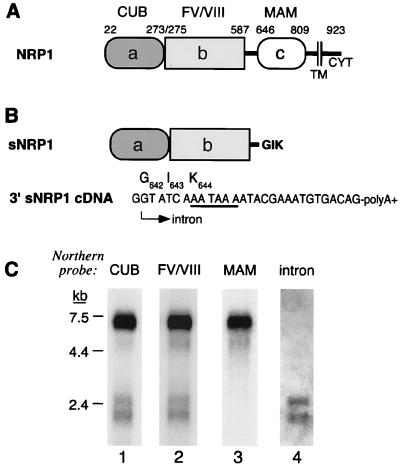
Figure 1.
sNRP1 structure and mRNA. (A) Schematic representation adapted from ref. 13 of NRP1. The a/CUB, b/factor V/VIII coagulation factor, and c/MAM homology domains as well as the transmembrane (TM) and cytoplasmic (CYT) domains are indicated. The number represents positions in the ORF corresponding to the boundaries of the various domains. (B) Schematic representation of sNRP1. The sNRP1 protein is identical to NRP1 up to Ser641, which is in the linker region between the b/coagulation and c/MAM homology domains and, in addition, contains three novel amino acids, G642 I643 K644. There is a unique, 28-bp, intron-derived sequence at the 3′ end of the cDNA. The 5′ end (arrow) of this sequence has a GT splice donor site, and its 3′ end is followed by a poly(A)+ tail. The cleavage/polyadenylation site (AATAAA), which contains a TAA stop codon, is underlined. The three new amino acids are shown above the codons. (C) Northern blot analysis of PC3 cell mRNA with probes corresponding to the a/CUB domain (lane 1), the b/coagulation factor domain (lane 2), the c/MAM domain (lane 3), and an oligonucleotide probe complementary to the sNRP1 28-bp, intron-derived sequence (lane 4). Each lane was loaded with 2 μg of PC3 mRNA.

