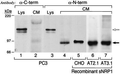
Figure 2.
Western blot analysis of sNRP1 protein expression. PC3 cell lysates (Lys) and CM were partially purified by Con A chromatography, resolved by SDS/PAGE, and probed with anti-NRP1 antibodies directed against the C-terminal (α-C-term) and N-terminal (α-N-term) regions of NRP1. Lanes: 1 and 3, PC3 cell lysates; 2 and 4, PC3 cell CM; 5, CHO cell CM from cells expressing recombinant His/Myc-tagged sNRP1; 6, AT2.1 cell CM from cells expressing recombinant sNRP1; 7, AT3.1 cell CM from cells expressing recombinant sNRP1. The open arrow indicates 130-kDa, full-length NRP1, and the solid arrow indicates 90-kDa sNRP1. The recombinant protein made by CHO cells is slightly larger because it is His- and Myc-tagged.

