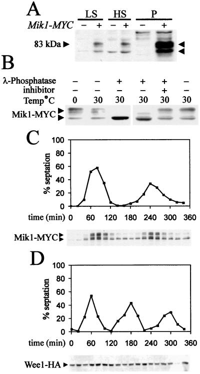
Figure 1.
(A) Equal amounts of total protein extracts from mik1-MYC (+) and untagged (−) cells grown to mid-log phase: low spin supernatant (LS), high spin supernatant (HS), and pellet (P). Arrowheads indicate bands specific to mik1-MYC extracts. (B) Mik1-MYC is resolved to a single form after phosphatase treatment. (C) mik1-MYC cells synchronized in G2 by elutriation. Progression through the cell cycle was followed by the septation index. Protein extract was prepared every 20 min, and 50 μg of total protein (pellet fraction) was analyzed. (D) An HA-tagged Wee1 strain was treated similarly.

