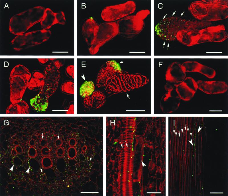
Figure 4.
Immunolocalization of the CN 8 antigen in cultured Zinnia cells (A–F), in the shoot apex of 14-day-old Zinnia seedlings (G and H), and in the stem of 2-month-old Zinnia plants (I). The yellowish green color indicates CN 8 epitope localization in the cell pseudocolored in red. Cells were cultured in the TE-inductive (A–E) or a control medium (F) for 30 h (A), 42 h (B), and 72 h (C–F) and treated with CN 8 scFv. (G) Cross section of midvein in a first foliage leaf. (H) Longitudinal section of midvein in a second foliage leaf. (I) Longitudinal section of stem vein. Arrows in C indicate the chloroplast-depleted area in an immature TE. The arrow and large and small arrowheads in E indicate a mature TE, a round CN 8-positive cell, and a semielongated CN 8-positive cell in which the CN 8 antigen localized in a tip, respectively. Arrows and large arrowheads in G–I and small arrowheads in G indicate mature TEs, immature TEs, and xylem parenchyma cells, respectively. (Bars in A–F = 30 μm; bars in G and H = 50 μm; bar in I = 100 μm.)

