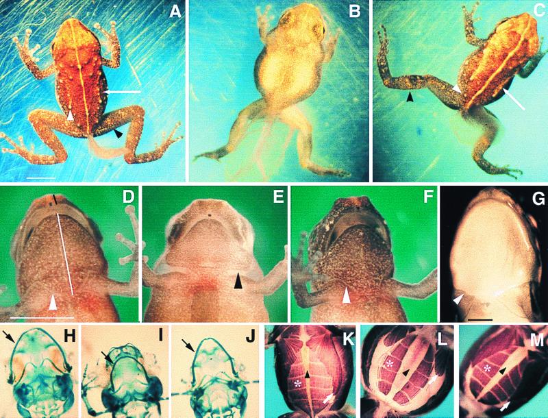
Figure 3.
Developmental effects of methimazole treatment. All E. coqui specimens are early posthatching stage. Scale bars represent 2 mm. (A) Control froglet with regressing tail. Adult skin characters include leg bands (black arrowhead), tubercles (white arrowhead), and raised dorsolateral ridges (white arrow). (B) Methimazole-treated sibling. (C) T3-rescued froglet. (D) Control froglet, ventral view. Skin has sutured (arrowhead). Black line indicates “snout–upper jaw” length, and white line represents the “lower jaw–suture line” length, measured for morphometric analysis (Table 2). (E) Methimazole-treated embryo, with a gap between head and trunk skin (arrowhead). (F) T3-rescued embryo. (G) Suture marks of newly metamorphosed Rana pipiens (arrowhead). (H–J) Meckel's cartilage (arrow) in control (H), methimazole-treated (I), and T3-rescued animals (J). (K) Control froglet, exhibiting convergence of the rectus abdominis (*) on the midline (arrowhead) and posterior elongation of the pectoral muscles (white arrow), events inhibited by methimazole (L) and rescued by T3 (M).

