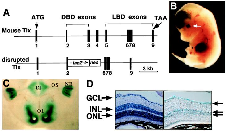
Figure 1.
Tlx expression in the mouse retina and optic stalk. (A) Schematic of the Tlx genetic locus (Upper) with predicted structure of the recombinant mutant allele (Lower). Exons are numbered. DBD, DNA binding domain; LBD, ligand binding domain. (B) Whole-mount LacZ staining of an embryonic day 13 heterozygous embryo showing Tlx expression in the neural retina, optic stalk, and forebrain. The arrow indicates the level of section shown in C. (C) DI, diencephalon; OS, optic stalk; NR, neural retina; OL, olfactory. (D) Retina sections of 3-week-old Tlx heterozygous mice have LacZ-positive cells (indicated by arrows) in the ganglion cell layer (GCL) as well as in the inner and outer nuclear layers (INL and ONL, respectively).

