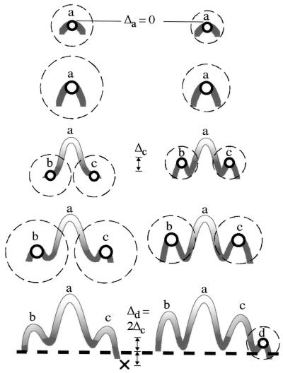
Figure 2.
Patterning cascade mode of tooth cusp development. The formation of each cusp begins by the appearance of the enamel knot (circle with thick line) that consists of nonproliferative epithelial cells expressing several molecular signals, including growth factors, and is surrounded by strongly proliferative epithelium (gray) and mesenchyme underneath (11). After the formation of the cusp tip, the enamel knot disappears and the epithelial and mesenchymal cells begin to differentiate into enamel-forming ameloblasts (lighter shading) and the dentin-forming odontoblasts (below the epithelium, not shown), respectively. The height difference between cusp a (Δa) of the left and right tooth is 0, but the subsequent enamel knots and cusps are at a distance that is determined by the previous enamel knot (hatched circle). This distance can be regulated by long-range inhibitors or by a decline in activators diffusing from the enamel knot (2, 11, 12). A small difference in the inhibitory field among teeth has a small effect in the position of the second set of cusps (c and b), but the effect is cumulative in the lower cusp d, resulting in teeth that are sharper (on the left) or blunter (on the right) in their cusp configurations. The actual number of cusps realized in each tooth crown is also determined by the termination of crown morphogenesis and the initiation of root formation (thick, hatched line). In the tooth on the left, the last developing cusp (d) would be initiated below the threshold and no cusp forms (marked with X). The anterior cusp b can have slightly different cusp spacing from the distal cusps. Note that the effects of inhibition and activation also can be realized by cell-to-cell-mediated propagation of the signal (13) and that both growth rate and the rate of spreading of the inhibitor can affect cusp spacing. Δa, Δc, and Δd denote the height differences between cusps a, c, and d, respectively, of the left- and right-hand side teeth.

