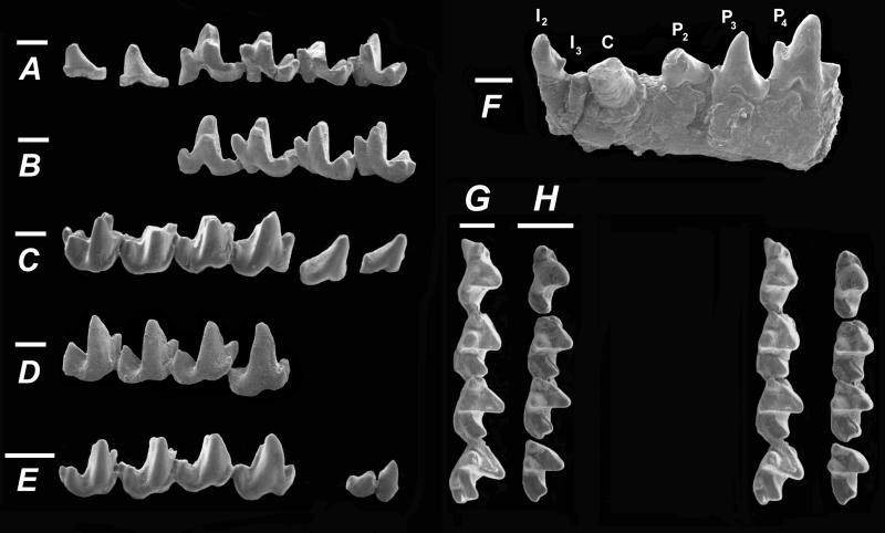
Figure 1.
Lingual views of DPC 15637, Widanelfarasia bowni, right P2–M3 (A), and cast of CGM 41878, W. bowni, left P4–M3 (reversed) (B). Labial views of DPC 15637, W. bowni, right P2–M3 (C); cast of CGM 41878, W. bowni, left P4–M3 (reversed) (D); and DPC 17427, W. rasmusseni, right P2 and P4–M3 (note that labial views are taken from slightly different orientations) (E). (F) Labial view of DPC 17779 (W. bowni), left dentary containing I2, root of I3, and C–P4. Note the loss of P1, the large cross-section and slightly procumbent orientation of the broken canine, and the enlarged I2 with posterior basal cusp. (G and H) Occlusal stereophotos of the holotypes of W. bowni (G, DPC 15637) and W. rasmusseni (H, DPC 17427). Note that a thin layer of matrix adheres to the hypoflexids of M1–M2 of DPC 17427. (Bars = 1 mm.)

