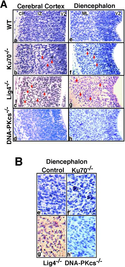
Figure 2.
Increased cell death in other subdivisions of developing CNS of Ku70−/− and Lig4−/− but not DNA-PKcs−/− embryos. (A) Hematoxylin- and eosin-stained sections (×400). Arrows point to the typical pyknotic cells, either isolated or clustered. (a–d) Cerebral cortex of E13.5 embryos (coronal sections). WT, wild type; CP, cortical plate. (e–h) Hypothalamic sulcus of the diencephalon of E13.5 embryos (transverse sections). (B) Enlarged images of Ae–Ah. Note pyknosis and hypocellularity in the ML of the diencephalon in the Ku70−/− (f′) and Lig4−/− (g′) embryos compared to DNA-PKcs−/− (h′) and control (e′) embryos. The darkly staining mitotic figures can be identified within the VZ of all control and mutant sections.

