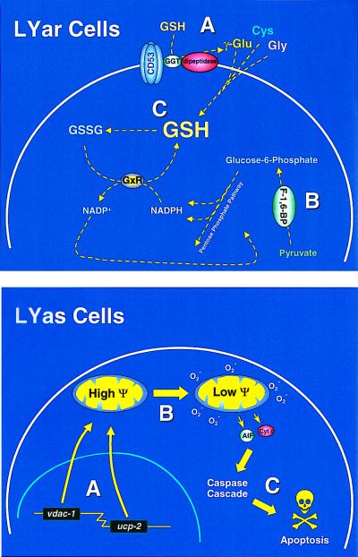
Figure 4.
Hypothetical LYar and LYas cells. From the data obtained, we have constructed a hypothetical model for the genes involved in apoptosis regulation. (Upper) LYar cells, which have previously been shown to have elevated GSH (C) (3), expressed message for CD53 and fructose-1,6-bisphosphatase that was not detected in the LYas cells. Both of these genes can combine to maintain elevated intracellular pools of GSH by enhancing the transport of GSH precursors into the cell (A) and keeping GSH in its reduced form by generating NADPH by enhanced gluconeogenesis and pentose phosphate pathway activity (B). GSSG, oxidized GSH; GxR, glutathione reductase. (Lower) In contrast, LYas cells trigger apoptosis in response to radiation by means of initiating transcription of proteins that adversely affect mitochondrial function. Radiation-induced increases in VDAC-1 and UCP-2 (A) uncouple mitochondrial electron transport and dissipate mitochondrial membrane potential (Ψ) (B), thus activating the release of apoptogenic factors. This leads to the activation of catabolic enzymes (primarily caspases), which complete the cell death process (C). AIF, apoptosis-initiating factor; Cyt c, cytochrome c.

