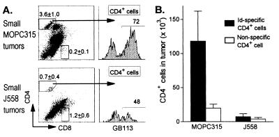
Figure 6.
Id-specific CD4+ cells accumulate in small Id+ tumors. Small MOPC315 and J558 tumors were triple-stained with the clonotype-specific GB113, anti-CD4, and anti-CD8 mAbs [MOPC315 (n = 5, 0.2 ± 0.1 g, 3.8 ± 0.6 × 10⁶ cells, serum M315: 29 ± 14 μg/ml); J558 (n = 4, 0.6 ± 0.4 g, 4.1 ± 2.8 cells, serum J558: 95 ± 54 μg/ml)]. (A) CD4 and CD8 cells were analyzed for TG TCR expression (GB113 mAb). The CD4 and CD8 gates were set on LN cells analyzed in parallel. A considerable number of tumor cells, outside the indicated CD4 and CD8 gates, stained variably for CD4 and CD8, but these cells were not bona fide T cells because they did not express TCR. By contrast, >80–90% of cells within the indicated CD4 gate were genuine T cells because they stained with the β-transgene (Vβ8)-specific F23.1 mAb (see Fig. 7A). Representative examples are shown; numbers represent mean frequencies ± SD. (B) Absolute number of Id-specific (GB113+) and nonspecific (GB113−) CD4+ cells in MOPC315 and J558 tumors.

