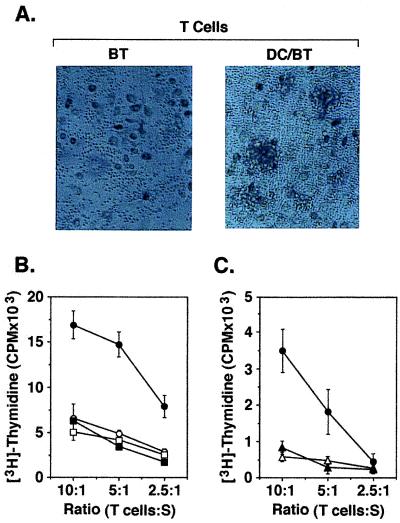
Figure 2.
Stimulation of T cells by DC/autologous breast tumor fusion cells. (A) T cells isolated from PBMC were incubated with autologous breast tumor cells (BT; Left) or autologous DC/breast tumor fusion cells (DC/BT; Right) at a ratio of 10:1 for 5 d in the presence of 20 units/ml HuIL-2. Incubation with the fusion but not the tumor cells resulted in the formation of T cell clusters. (B) T cells were cultured with autologous DCs (○), autologous breast tumor cells (□), autologous breast tumor cells mixed with DCs (■), or autologous breast tumor/DC fusion cells (●) at the indicated ratios of T cells to stimulators (S). (C) T cells were cultured with polyethylene glycol-treated autologous DCs (▵), autologous DCs fused to autologous monocytes (▴), or autologous breast tumor/DC fusion cells (●) at the indicated ratios. After 7 d, uptake of [3H]thymidine was measured during a 12-h incubation. The results are expressed as mean ± SD of three replicates.

