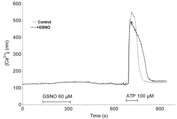
Figure 5.
Plot of the intracellular calcium concentration in the CFBE cells. In the control experiment (continuous line) the cells were exposed only to ATP (the duration is indicated by the horizontal line). In another experiment (dotted line) the cells were exposed first to GSNO, then, after a 5 minutes washing step, to ATP. The two results were overlapped. Plots represent the averaged results of 3–6 experiments.

