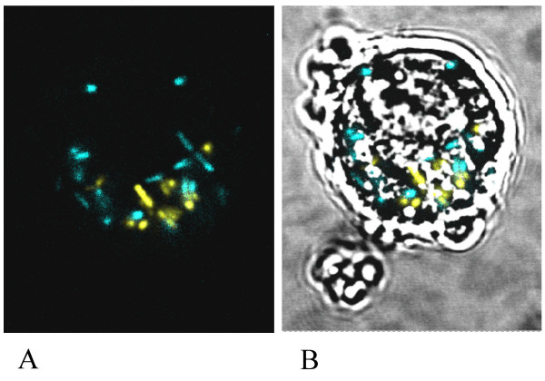
Figure 6.

Visualization of internalized bacteria. Caco-2 cell containing a mixture of blue/cyan wt-CFP and yellow wt-YFP L. monocytogenes. A: Fluorescence microscopy image. B: Overlay of fluorescent image and phase contrast image showing the Caco-2 cell.
