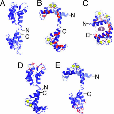
Fig. 2.
Conformations of WT CaM and the designed mutant CaMs. (A) NMR structure of Ca2+-free CaM (PDB ID code 1CFD) (33). (B) X-ray crystal structure of Ca2+-bound CaM (PDB ID code 3CLN) (34). The hydrophobic patches important for target recognition are shown in red. Ca2+ atoms are shown as yellow spheres. (C) X-ray crystal structure of Ca2+/CaM bound to CaMKII-cbp, shown in pink (PDB ID code 1CM1) (35). (D and E) Models of the mutant CaMs designed to bind Ca2+ only in the C-terminal domain, CaM-CWT (D), and only in the N-terminal domain, CaM-NWT (E). These models were generated by combining the structure of the WT Ca2+/CaM domain with the structure of Ca2+-free CaM representing the mutated CaM domain. Mutant residues predicted by the computation to produce the best stabilized structure (Table 1) are shown in red. The figure was generated with PyMOL (DeLano Scientific, South San Francisco, CA).

