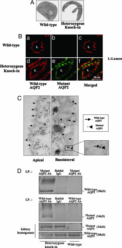
Fig. 3.
Renal morphology and localization of wild-type and mutant AQP2 in the collecting duct cells. (A) Kidneys from wild-type and mutant AQP2 knockin mice at 8 weeks of age. Polyuria-induced hydronephrosis was observed in mutant mice. (B) Double immunofluorescence of wild-type and mutant AQP2 protein in the collecting ducts of wild-type and heterozygous mutant mice. (a–c) In wild-type mice, wild-type AQP2 was localized to the apical membrane after dehydration. (d–f) Mutant AQP2 was localized to the basolateral membrane. Similarly, wild-type AQP2 is recruited to the basolateral membrane, although some wild-type AQP2 remained in the apical membrane. (C) Double-immunogold staining of wild-type and mutant AQP2. Arrows and arrowheads indicate wild-type and mutant AQP2, respectively. Little apical localization of mutant AQP2 and basolateral localization of wild-type AQP2 were seen. (D) Coimmunoprecipitation experiments of wild-type and mutant AQP2. Homogenates of kidney papillae from wild-type mouse and heterozygous knockin mouse were immunoprecipitated with mutant AQP2 antibody (Top) or wild-type AQP2 antibody (second image from the top). Rabbit IgG was also used as a negative control for immunoprecipitation. Samples immunoprecipitated with wild-type AQP2 were probed with anti-mutant AQP2 antibody and vice versa.

