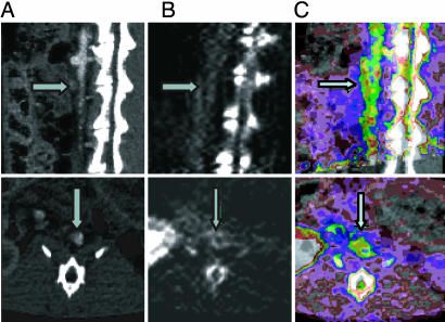
Fig. 4.
Coregistration of CT and PET images. CT images (A) and PET images (B) were coregistered (C) by employing a workstation (REVEAL-MVS) that enables multimodal standard (rigid) image fusion using anatomic landmarks. After PET scanning, CT angiography was performed with a Siemens Medical Solutions SOMATOM Sensation 16-slice CT scanner (Siemens Systems, Forcheim, Germany). Three-dimensional, multiplanar reconstruction of raw images was performed, and the CT images were then coregistered with the PET images. With the CT/PET image as a guide, region-of-interest segments of the thoracic and abdominal aorta were drawn. Accumulation of radioactivity was calculated for each region of interest and expressed as percentage injected dose per milliliter. A value of P < 0.05 was considered significant. Upper row, sagittal sections; Lower row, transverse sections. Arrows in A indicate lumen with CT contrast; arrows in B indicate arterial wall plaque; and arrows in C show the fusion of lumen CT contrast in wall plaque in the PET image.

