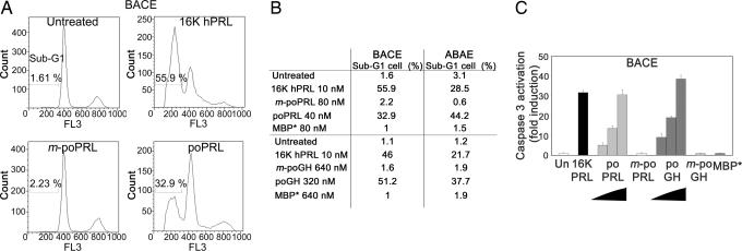
Fig. 2.
The tilted peptides of hPRL and hGH induce apoptosis of BACE and ABAE cells by activating caspase-3. (A and B) poPRL and poGH induce endothelial cell apoptosis. FL3, red fluorescence intensity. (B) Percentage of cells entering apoptosis (sub-G1 population). BACE (A and B) or ABAE (B) cells were treated with 10 nM 16K hPRL or with a fusion protein for 18 h. Cell cycle progression was monitored by measuring cell DNA content by flow cytometry analysis. (C) poPRL and poGH activate caspase-3 in endothelial cells. poPRL (10, 20, and 40 nM), poGH (80, 160, and 320 nM), or 10 nM 16K hPRL induced caspase-3 activation in endothelial cells as compared with untreated cells (Un). Control proteins, i.e., 80 nM mutated poPRL (m-poPRL), 640 nM mutated poGH (m-poGH), and 640 nM MBP* did not. BACE cells were treated for 18 h with the indicated proteins. Each result is expressed as an enhancement factor (treated vs. untreated cells). Each bar represents the mean ± SD; n = 3.

