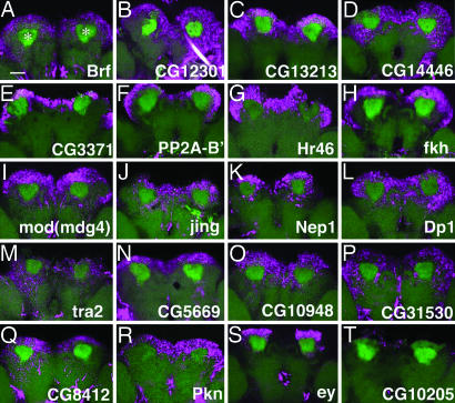
Fig. 2.
In situ hybridization of MB genes in the adult brain. (A–T) Expression patterns of MB genes (magenta) in the adult brain as revealed by fluorescent in situ hybridization. Reconstruction of confocal images of the posterior part of the brain. MBs are visualized with UAS-mCD8::GFP driven by OK107-Gal4 (green). Note that the specific GFP signals were compromised during hybridization, and other neuropil structures are also visible by nonspecific green fluorescence. Asterisks in A indicate the MB calyces. (Scale bar, 50 μm.) CG10205 is included as a negative control (belonging to rank-D in Table 3).

