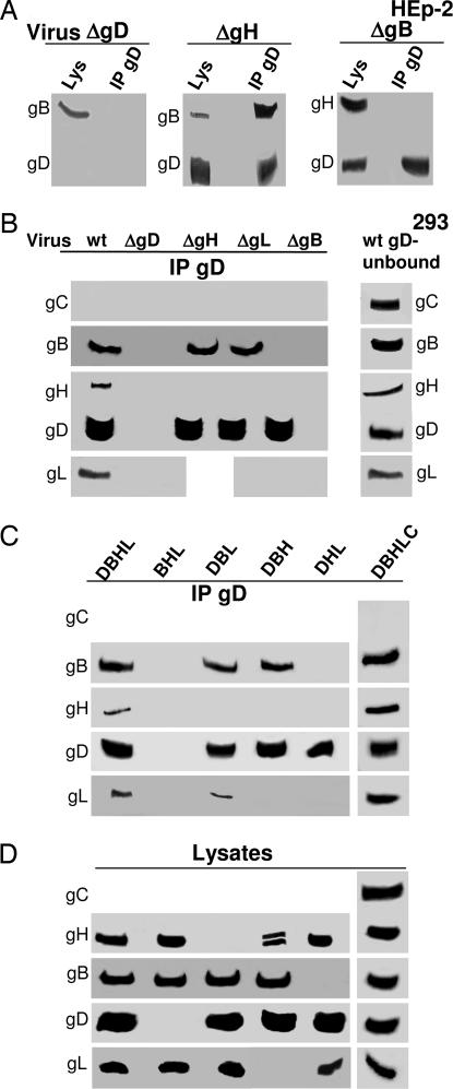
Fig. 6.
Analysis of immuno- and coimmunoprecipitated proteins from cells infected with deletion viruses or transfected with the indicated mixtures. (A) HEp-2 cells. (B–D) 293 cells. (A and B) Cells infected with WT virions (wt) or ΔgD, ΔgB, ΔgH, and ΔgL viruses as gD−/+, gB−/+, gH−/+, and gL−/+ virions. (C and D) 293 cells transfected with the indicated mixtures containing the plasmids encoding for gD, gB, gH·gL, gC, and twice the amount of HVEM plasmid. DBHL, gD, gB, gH, gL; BHL, gB, gH, gL; DBL, gD, gB, gL; DBH, gD, gB, gH; DHL, gD, gH, gL; DBHLC, gD, gB, gH, gL, gC. In all panels, gD was immunoprecipitated with pAb R8 to gD. Immuno- and coimmunoprecipitated proteins were analyzed by WB. (D) Analysis of cell lysates from the experiment shown in C.

