Fig. 2.
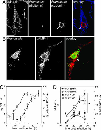
Intracellular Francisella become enclosed in large vacuoles after intracytoplasmic replication. (A) Confocal micrographs of an LVS-infected BMM at 24 h p.i., subjected to the phagosomal integrity assay. Cytoplasmic bacteria (red and green, appearing yellow in the overlay) are labeled after digitonin permeabilization, whereas clustered bacteria are detected only after saponin permeabilization (red). Calnexin staining (blue) allows detection of digitonin-permeabilized cells. (B) Confocal micrographs of an LVS-infected BMM at 24 h p.i. BMMs were infected with LVS, fixed, and processed for immunofluorescence with Francisella LPS and LAMP-1 antibodies. Bacterial clusters (green) are enclosed in LAMP-1-positive, membrane-bound compartments (red) termed Francisella-containing vacuoles (FCVs). Arrows indicate FCVs. (Scale bars: 10 μm.) (C) Kinetics of intracellular replication and FCV formation. BMMs were infected with LVS for the indicated times. Intracellular bacteria were enumerated from cfus, and FCV formation was measured as the percentage of infected cells harboring LAMP-1-positive FCVs. (D) Effect of inhibition of bacterial protein synthesis on FCV formation and replication. BMMs were infected with LVS and left untreated or treated at 14 h p.i. with 10 μg/ml chloramphenicol (indicated by arrow), and FCV formation (filled shapes) or intracellular growth (open shapes; cfu) was measured. The asterisk indicates statistically significant differences between control and chloramphenicol-treated BMMs at 28 h p.i. (P < 0.05, two-tailed unpaired Student’s t test).

