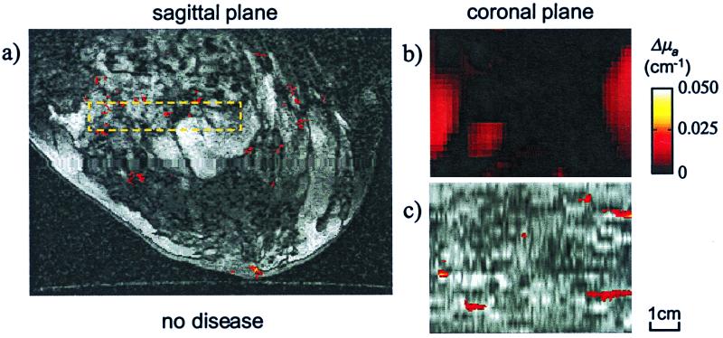
Figure 5.
Case III: No disease. (a) Functional sagittal MR image after Gd contrast enhancement passing through the middle plane of the breast. (b) Coronal DOT image, perpendicular to the plane of the MRI image in a, for the VOI indicated on a with the interrupted line box. (c) Functional MR coronal reslicing of the VOI with the same dimensions as b.

