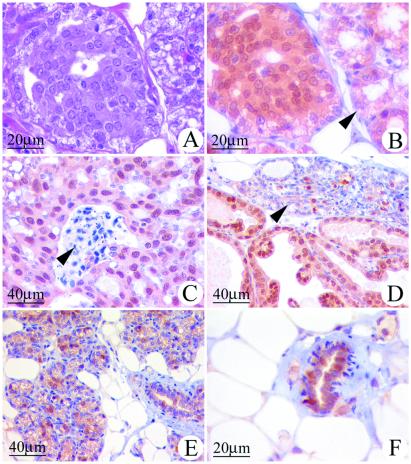
Figure 6.
Atypical epithelial hyperplasias and mammary cancers induced in ACI rats by E2 express PR. Cells expressing PR were identified immunohistochemically as described in Materials and Methods. (A) Mammary gland, sectioned and stained with hematoxylin and eosin, from a 21-wk-old female ACI rat treated with E2 for 12 wk. Illustrated is a focal region of atypical epithelial hyperplasia and, to the right, adjacent acinar structures. The epithelial cells within the atypical hyperplasia exhibited enlarged nuclei and dense eosinophilic cytoplasmic staining. The illustrated atypical hyperplasia was minimally deviated relative to the surrounding lobules. The number of atypical hyperplastic foci and the degree of cellular atypia were observed to increase as a function of the duration of E2 treatment beyond 12 wk (data not illustrated). (B) A serial section to that in A that has been immunostained for PR. The majority of cells within the atypical epithelial hyperplasia exhibited immunoreactivity to PR, whereas fewer cells in the adjacent acinar structures stained positive for PR (arrow). (C) Mammary comedo carcinoma from an ACI rat treated with E2 for 193 days. The majority of the cancer cells exhibited immunoreactivity to PR. The arrow indicates necrotic debris characteristic of these cancers. (D) Mammary papillary carcinoma from an ACI rat treated with E2 for 216 days. The majority of the epithelial cells within the cancer exhibited immunoreactivity to PR. The arrow indicates adjacent acinar structures where a subset of the epithelial cells were immunoreactive to PR. (E) Mammary gland from an ACI rat treated with E2 for 12 wk exhibited lobuloalveolar hyperplasia. A subset of the epithelial cells stained positive for PR. (F) Mammary gland from an untreated, 21-wk-old, ovary-intact ACI rat; age-matched control for the E2-treated animals illustrated in A, B, and E. A subset of epithelial cells within the normal ductal structures exhibited immunoreactivity to PR.

