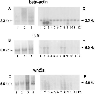
Figure 2.
Expression of fz5, wnt5a, and β-actin in RA tissue samples and adult tissues by Northern analysis. A–C show the expression of β-actin, fz5, and wnt5a in RA tissue specimens. D–F show the expression of the same genes in 12 adult tissues that are peripheral blood leukocyte, lung, placenta, small intestine, liver, kidney, spleen, thymus, colon, skeletal muscle, heart, and brain (lanes 1–12, respectively, in D–F). (A) Northern blot showing 2.3-kb β-actin-specific band in three RA tissue specimens; (B) 5-kb fz5-specific band in three RA tissue specimens; (C) lanes 1–3, 5-kb wnt5a-specific band in three RA specimens; lane 4, wnt5a-specific band in fetal fibroblast as a positive control. (D) Northern blot showing 2.3-kb β-actin-specific band in 12 different adult tissue specimens; (E) 5-kb fz5-specific band in the 12 adult tissue Northern blot; (F) result of wnt5a probe hybridization by using the same Northern blot. Specific activity of the probes used for hybridization and RNA concentrations of the tissue samples was the same for each analysis. The adult multiple tissue Northern blot was stripped and reprobed during the course of the experiments, and β-actin hybridization was performed last of all.

