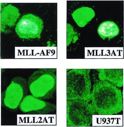
Figure 2.
Immunofluorescent staining of U937T cells expressing MLL-AF9 and deletion mutants MLL3AT and MLL2AT, as well as the U937T parent. Cells were induced by tetracycline withdrawal for 48 hr and were stained with M2 FLAG monoclonal antibody followed by goat anti-mouse Alexa 488 conjugate. MLL-AF9 and MLL3AT have a predominantly punctate subnuclear distribution (green background with bright green/white spots), whereas MLL2AT is uniformly distributed in the nucleus (uniform green nuclear staining). (×1,000.)

