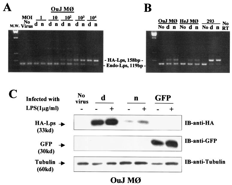
Figure 4.
Evidence of gene transfer and expression in transduced C3H/HeOuJ macrophages. (A) The result of PCR on DNA extracted from peritoneal macrophages of C3H/HeOuJ mice primed i.p. with 3 ml of 3% thioglycollate solution (Sigma) 3 days before cell collection. The amount of genomic DNA used in each lane was estimated to be equivalent to 4,000 cells. OuJ Mφ represents macrophages from C3H/HeOuJ mice. (B) The result of RT-PCR on C3H/HeOuJ macrophages after adenoviral gene transfer. HeJ Mφ represents macrophages from C3H/HeJ mice; 293 represents human embryonic kidney 293 cells. (C) Western blot analysis on lysates from C3H/HeOuJ cells after adenoviral gene transfer. d, Ad5-Lpsd/Ran virus; n, Ad5-Lpsn/Ran virus; no, no virus; IB, immunoblot.

