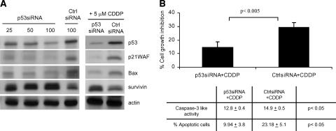
Figure 4.
p53-specific death is induced by CDDP in MPM cells. (A) ZL34 cells were transfected with the indicated concentrations of p53 siRNA or control siRNA for 24 hours, or were treated with 50 nM p53 or control siRNA for 24 hours and incubated with 5 µM CDDP for a further 48 hours. Lysates were analyzed by immunoblotting using antibodies specific for p53, p21WAF, Bax, and survivin. Staining of actin was used as loading control. (B) ZL34 cells were transfected with 50 nM p53 siRNA or control siRNA for 24 hours and were subsequently incubated for 48 hours with 5 µM CDDP. Cell growth inhibition was calculated as the percentage relative to untreated controls. Representative data of three independent experiments are shown. Caspase-3-like activity and the percentage of apoptotic cells were determined by Annexin V staining and flow cytometry, as described in Figure 3. Student's t test was used to determine the significance between the p53 siRNA + CDDP group and the control siRNA + CDDP group.

