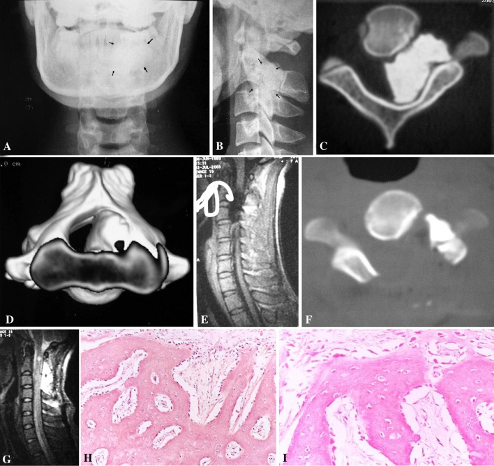
Fig. 1.
a Cervical A–P view X-ray showed densification at left side at C2–3 level. b Cervical lateral view X-ray showed high density at C2–3 level, the facet joints between C2\C3 were obscure. c CT scan through the intervertebral foramen between C2 and C3 showed the tumor occupied more than 60% canal and the vertebral shape changed, caused by the chronic pressure. d Three dimensions CT reconstruction showed the three-dimension relationship between the tumor and the cervical spinal canal. e Middle sagittal plane T1WI MRI showed the spinal cord was pressed by the tumor. The signal of the tumor was low. f The same plane as c after operation, showed the whole decompression of the spinal canal, part of the tumor, near the vertebral artery was left. g The same plane as e after operation, showed the whole decompression of the cord and spinal canal. h, i The pathological study showed that the lesion was composed of uniformly dense, compact, cortical-like mature lamellar bone. H: ×100, I: ×400

