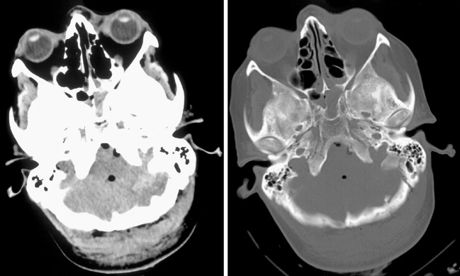
Fig. 1.
Axial cranial CT scans (corresponding soft-tissue and bone window setting) revealing severe head injury accompanied by traumatic internal pneumocephalus with air distributed prepontine, perimesencephally and intraventricullary accompanied by skull fractures of the sphenoid, left occipital and petrosal bone leading to PR

