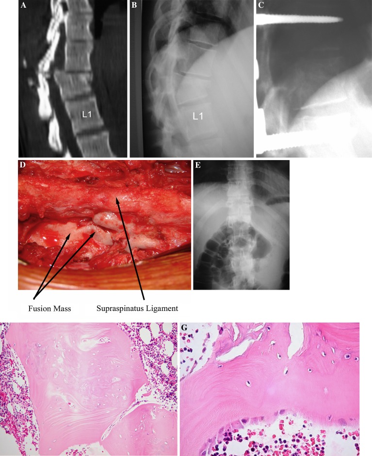
Fig. 1.
This 30-year-old female sustained an AO type C2.1 fracture of T12 after a fall from a horse. Initial work-up in the ER did not show any other lesion. She was neurologically intact. We performed a T11-L1 posterior short segment fusion as previously described. Implant removal was performed 14 months following the index surgery. The biopsy demonstrated irregular bony trabeculae, foci of enchondral ossification and numerous osteoblasts, all characteristic elements of ongoing bone remodeling. Several aggregates of lymphoplasmocytes were observed and have been attributed to a foreign body inflammatory type of response. a Preoperative sagittal CT. b Preoperative lateral radiograph. c Lateral radiograph at 5 months postop. d Perioperative image of extensive new bone growth during the secondary surgery. e AP radiograph 15 months following the index surgery after implant removal. f Histology at 100× magnification. g Histology at 400× magnification

