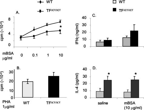
Figure 4.
mBSA-specific spleen T-cell proliferation. Spleen cells were cultured in the absence or presence of the indicated amount of mBSA (A) and phytohemagglutinin (B) for 48 hours. Cultures were pulsed with [3H]-thymidine for the final 18 hours and the incorporated radioactivity was measured. Results are expressed as means ± SEM of eight mice in each group [*, P < 0.05 for TFδCT/δCT mice versus TF+/+ (WT) controls]. mBSA-specific T-cell cytokine expression. Spleen cells were cultured in the absence or presence of mBSA for 48 hours. Supernatants were analyzed by ELISA for IFN-γ (C) and IL-4 (D). Results are expressed as means ± SEM of eight mice in each group (*, P < 0.005 for vehicle versus mBSA treatment).

