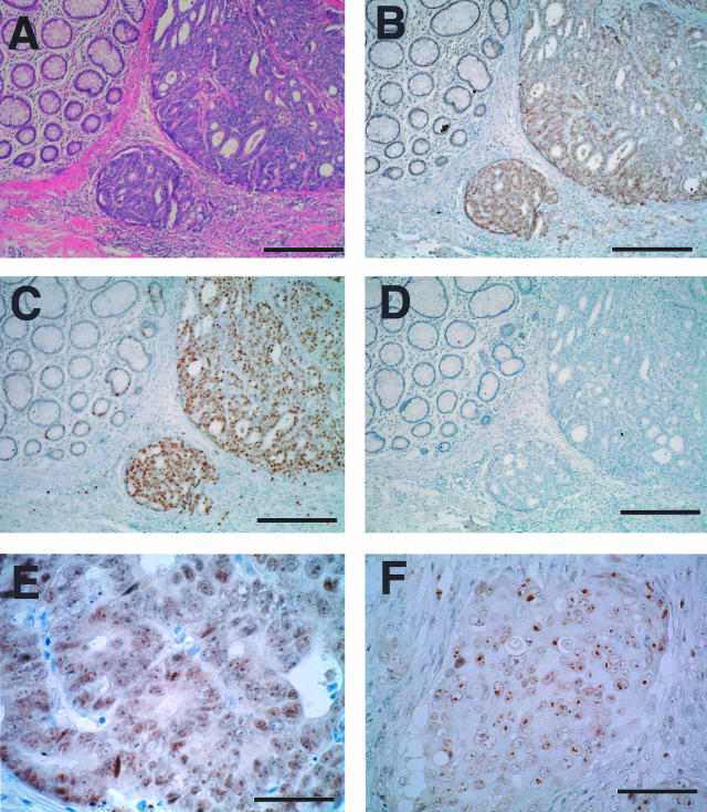
Figure 3.
Immunohistochemical staining of Mina53 and Ki-67 proteins in colon tumor tissues. A: H&E staining of a section that contained a moderately differentiated adenocarcinoma. B: Serial section of A stained by anti-Mina53 antibody showing elevated expression of Mina53 in the tumor area but little staining in the adjacent nonneoplastic tissue. C: Serial section of A stained by anti-Ki-67 antibody. D: Control section of A in which the primary antibody was omitted. E: A high-power field of the section shown in B showed the characteristic nuclear localization of Mina53. F: Mina53 staining of a poorly differentiated adenocarcinoma. Scale bars: 300 μm (A–D); 50 μm (E); 75 μm (F).

