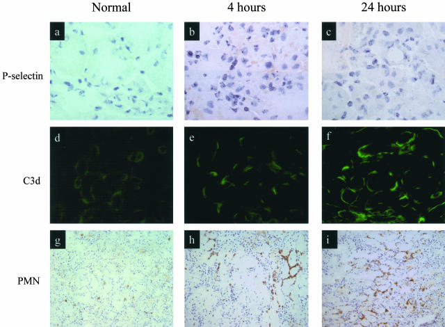
Figure 2.
Staining for P-selectin expression, complement activation product, and neutrophil infiltration. Normal untreated WT mice and WT mice treated with 55 minutes of bilateral renal ischemia followed by reperfusion for 4 and 24 hours. a, b, and c: P-selectin expression is seen on corticomedullary small peritubular vessels. d, e, and f: Immunochemical staining of C3d is present on the basolateral surface of renal tubular epithelium. g, h, and i: Immunochemical staining of neutrophils in the corticomedullary junction. Original magnifications: ×1000 (a–c); ×400 (d–f); ×250 (g–i).

