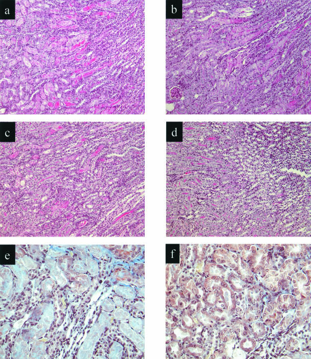
Figure 4.
Effect of renal ischemia and reperfusion on renal morphology and fibrin deposition in WT and C3-deficient mice pretreated with anti-P-selectin or control mAb. Light microscopy of kidney corticomedullary junction showing tubular damage at 48 hours of reperfusion. a: WT control-treated; b: WT anti-P-selectin-treated; c: C3-deficient control-treated; d: C3-deficient, anti-P-selectin-treated mice. e and f: Martius scarlet and blue staining at damaged tubular area in WT control-treated (e) and C3-deficient, anti-P-selectin-treated (f) mice. Fibrin, erythrocytes, nuclei, and connective tissue stain as red, yellow, black, and blue, respectively. Original magnifications: ×250 (a–d); ×400 (e, f).

