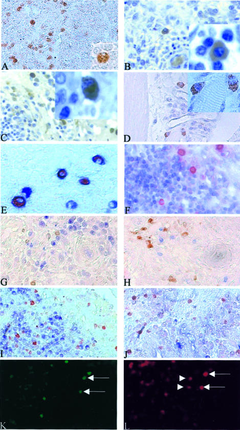
Figure 4.
In normal activated lymph nodes, RA synovium, and DM, Th1-producing cells presented a plasma cell-like morphology. In RA synovium, these Th1 plasmacytoid-like-producing cells still express the CD4 but not the CD3 and B/plasma cell markers. In RA synovium, some of the IFN-γ+ cells can express IL-17. Immunostaining using anti-IL-17 and anti-IFN-γ antibodies (DAB, brown) were performed on normal activated lymph nodes, RA synovium, and DM paraffin-embedded sections. In activated lymph nodes, IL-17 or IFN-γ+ cells detected in the T-cell zone out of germinal center (A) have a plasma cell-like morphology (A, inset). In RA synovium, the IL-17- and IFN-γ-producing cells presented the same morphology (insets of B and C, respectively). IL-17+ cells and IFN-γ+ cells were either isolated (B) or part of a lymphocytic infiltrate (C). In DM-, IL-17-, or IFN-γ-producing cells detected in lymphocytic infiltrates in the endomysium (D) presented also a plasma cell-like morphology (D, inset). Double staining using anti-IL-17 or anti-IFN-γ (AEC, red) and anti-CD4 or anti-CD3 antibodies (Vector blue, blue) were performed on RA sections. IL-17+ cells express the CD4 marker (E) but not the CD3 (F). Double staining for IL-17 or IFN-γ (red) and CD20, CD38, κ or λ light chains (blue) showed that none of the IL-17- or IFN-γ-positive cells was expressing CD20, CD38, κ or λ light chains (G to J, respectively). Same sections of RA synovial tissue were incubated with anti-IL-17 and anti-IFN-γ and stained using the immunofluorescence technique (K and L, respectively). Some cells could be expressing at the same time both IL-17 (fluorochrome fluorescein isothiocyanate, green) and IFN-γ (fluorochrome CyaninIII, red) as indicated by the arrows in K and L. These experiments also showed that some cells secreting IFN-γ were not secreting IL-17 as indicated by the arrowheads in L. Original magnifications: ×400 (A–D, F–L); ×1000 (E and insets in A–D).

