Figure 1.
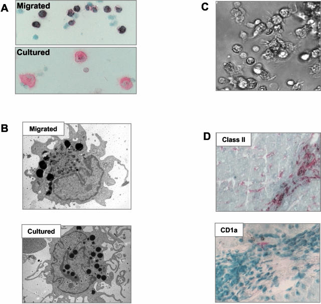
Appearance of cultured liver DCs. A: Cells isolated after migration and stained using APAAP for class II expression, have a monocyte-like morphology. After further culture the cells acquire a more typical DC appearance. B: Transmission electron microscopy of DCs after overnight migration and after 2 days further culture. The migrating cells have an oval nucleus and dense cytoplasm, many of the cells had very dark staining vesicles in the cytoplasm. The cultured matured cells had a more complex structure of dendrites or veils and a more open and lobed nucleus. C: Cells migrated from normal liver and cultured for 2 to 3 days develop characteristic veils and a typical DC morphology. D: Class II-positive cells in the portal tract of normal liver have the appearance of DCs but are CD1a-negative. CD1a is not detected in normal liver and only very occasional CD1a cells are detected in inflamed liver (liver tissue is from a patient with chronic liver allograft rejection). Original magnifications: ×20 [A, D (bottom)]; ×5000 (B); ×10 (D, top).

