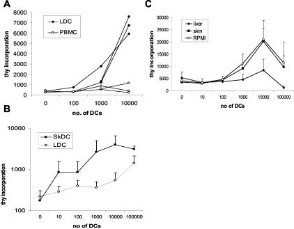
Figure 3.
Capacity of liver DCs to induce proliferation of allogeneic T cells. A: Migrated liver-derived DCs stimulate naïve T-cell proliferation. Proliferation of T cells in response to liver-derived DCs was measured in a thymidine incorporation assay. Varying numbers of stimulator cells, liver DCs, or PBMCs, were used to stimulate 2 × 10⁵ cord blood mononuclear cells in a 5-day assay. Mean of triplicate wells is shown. Each line represents a different experiment with different DC preparations (n = 3). B: The ability of DCs from skin (n = 3) and liver (n = 5) to stimulate allogeneic T cells was tested in a modified MLR in which highly purified, negatively selected allogeneic T cells were used as responder cells and DCs that had been purified using negative selection as stimulators. The skin DCs were significantly better stimulators of allogeneic T cells than liver-derived DCs (P < 0.0001). C: The influence of soluble tissue factors to modify the response of DCs after overnight migration was tested using the media from these cultures. mDCs were cultured overnight in tissue-conditioned media or complete media, before comparison in a modified MLR using pure adult allogeneic T cells (n = 4). mDCs cultured overnight in liver tissue-conditioned media were poor stimulators of T-cell proliferation compared to normal mDCs (P < 0.001).

