Figure 10.
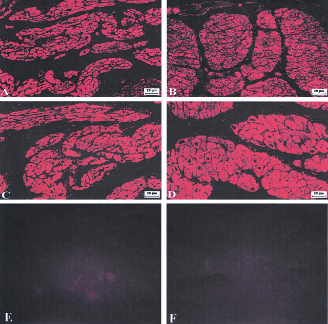
Immunolocalization of CaD and α-actin in control and obstructed bladders. Semithin (3 μm) cross-sections were prepared from normal and obstructed bladders and probed by indirect immunofluorescence using anti-caldesmon, anti-α actin antibodies (1:250), and CY3-conjugated anti-mouse IgG. Photographs were obtained under a fluorescence microscope. The smooth muscle layers from normal (A) and decompensated (B) bladders reacted with anti-caldesmon antibody. Adjacent sections from both bladders were reacted with anti-actin antibody (C, normal bladder and D, decompensated bladder). Notice that the immunostaining of the muscle bundles using α-actin and CaD antibodies are similar. E and F are negative controls for CaD and α-actin, respectively, carried out on adjacent sections from decompensated bladder, treated similar to A–D except that the anti-CaD and anti-α-actin antibodies were replaced by normal mouse serum. Magnifications are shown by the bars in the photographs.

