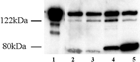
Figure 8.
Western blot analysis of h-CaD and l-CaD in different bladder groups. Western blots revealing h-CaD and l-CaD as detected by anti-caldesmon antibody. Lane 1, purified chicken gizzard h-CaD; lane 2, control bladder; lane 3, sham-operated bladder; lane 4, compensated bladder; lane 5, decompensated bladder. The small bands below the h-CaD band in well 1 are proteolytic fragments, exaggerated by overloading of the well by purified h-CaD.

